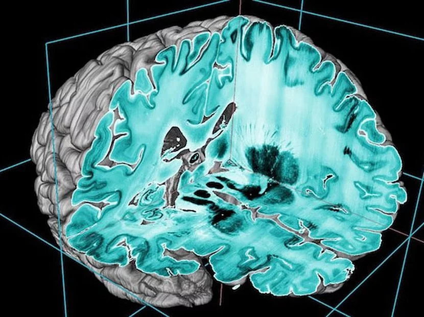
MRI (magnetic resonance imaging) has become the method of choice for examining the central nervous system (CNS), the brain and spinal cord. MRI provides unparalleled detail resolution of all complex structures of the CNS. It enables the differentiation of gray and white brain matter, cranial nerve nuclei and axonal nerve cords, cranial nerves, vessels, cerebrospinal fluid and meninges.
Our recommendation
An MRI scan of the brain can detect tumors, inflammation, strokes and other neurological diseases at an early stage.

MRI is primarily used in the area of the brain and spinal cord to determine:
- Brain tumors: glioblastoma, meningioma, etc.
- Vascular malformations: Aneurysms, angiomas, etc.
- Circulatory disorders: Micro- and macroangiopathy, vascular occlusion, stroke, encephalopathy in hypertension, etc.
- Inflammatory and infectious brain diseases: Multiple sclerosis, meningitis, etc.
- Neurodegenerative diseases: Alzheimer’s disease, Parkinson’s disease, etc.
Stroke diagnostics with diffusion-weighted MRI
Strokes are characterized by the sudden loss of brain function: Acute weakness or paralysis on one side of the body, numbness, speech difficulties, visual disturbances and dizziness or even unconsciousness, as well as sudden severe headaches and nausea. These symptoms are triggered by circulatory disorders of the brain, usually caused by vascular occlusion. This can lead to bleeding in the brain.
If these or similar symptoms occur, it is important to act quickly. This is because there is only a short window of opportunity to treat the circulatory disorder and save the affected regions of the brain.


Diffusion Weighted Imaging (DWI)
Determination of the risk of stroke
Most strokes are avoidable. They are caused by deposits in the heart, the main artery (aorta) and the arteries supplying the brain. They usually announce themselves through minor strokes or so-called transient ischemic attacks (TIAs), which are then followed by a massive stroke that leads to paralysis, loss of speech or death. Examination of the brain with MRI can visualize the precursors of a stroke – small changes in the brain substance that can occur unnoticed.
Examination of the heart with cardiac CT/MRI and MR angiography and of the aorta and the arteries supplying the brain. carotid and vertebral arteries can detect a risk of stroke at an early stage. Although ultrasound examinations are often used for this purpose, they are in the area of the left atrial appendage, where most of the thrombi that trigger strokes form.
Get advice. A stroke is preventable. Strokes can be prevented by recognizing the warning signs early and treating them with medication.
Even today, if a stroke is suspected, a computer tomography (CT) scan of the brain is often carried out – to rule out a hemorrhage. However, CT does not show the actual infarction of the brain in the early stages.
Diffusion-weighted MRI, on the other hand, can reliably detect a stroke just minutes after the onset of the circulatory disorder – early enough to initiate targeted treatment.
Important to know: strokes often announce themselves. In one in three patients, the “major” stroke is preceded by smaller, so-called “transient ischemic attacks” – TIAs – with similar symptoms such as numbness or paralysis in one half of the body or sudden speech and vision problems. However, these disappear on their own within 24 hours. TIAs can also be detected with diffusion-weighted MRI – so that the “big”, irreversible stroke can be prevented.
Intracranial vessels
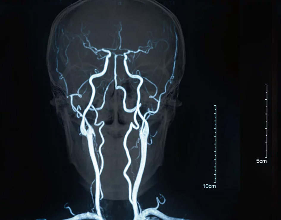
The visualization of intracranial vessels by CT or MR angiography plays an important role in circulatory disorders of the brain, but also in vascular malformations, e.g. aneurysms, arteriovenous shunts or angiomas.
Depending on the problem, computed tomography (CT) or magnetic resonance imaging (MRI) angiography (vascular imaging) offers advantages.
MR angiography of the intracranial vessels is usually performed without contrast medium. It is radiation-free and also allows the brain tissue to be assessed in a single examination.
An MR angiography can save lives
Early detection of aneurysms
It is therefore important to detect aneurysms at an early stage. Since headaches can be an early sign of cerebral aneurysms, an MR angiography should be performed if headaches occur. Although aneurysms are a rare cause of headaches, the consequences of an overlooked aneurysm are catastrophic.
The illustration on the left shows an anerysm from the outside. It was clipped: In the area where the aneurysm leaves the aneurysm, the aneurysm neck, a small clip was applied, which closes the aneurysm and cuts it off from the blood flow. A rupture is no longer to be feared.
The image on the right shows a cross-section of an aneurysm that has been “coiled”: A wire mesh was first inserted into the aneurysm via a fine catheter (green), which causes the blood to clot and thus seals the aneurysm from the inside. A stent (vascular support made of wire mesh) ensures that the coils remain in the aneurysm.
If an aneurysm is detected, it can be closed by means of neurosurgery or a neuroradiological catheter procedure. The aneurysm is surgically “clipped” by applying a small clip to the neck of the aneurysm. Without open surgery, aneurysms can also be treated using a catheter, a thin tube that is inserted into the brain via the inguinal artery. This involves inserting material (coils) into the aneurysm via the catheter, which leads to blood clotting in the aneurysm (thrombosis) and closes the aneurysm in this way – this is known as “coiling”.
Examination of Family Members in the Event of an Aneurysm Diagnosis
Data from a large study on familial aneurysms, the Familial Intracranial Aneurysm Study, shows that family members of patients with an aneurysm have a significant risk of also developing an aneurysm. For first-degree relatives (parents, children and siblings), the probability is around 20%. Women and people who have smoked in the past and/or have high blood pressure are particularly at risk.
Therefore, aneurysm screening with an imaging examination of the cerebral arteries is usually recommended, especially in first-degree relatives. If an aneurysm is found, a neurosurgeon must decide whether treatment of the aneurysm is necessary. If no aneurysm is detected, regular repeat examinations should be carried out, as an aneurysm may develop over time.


