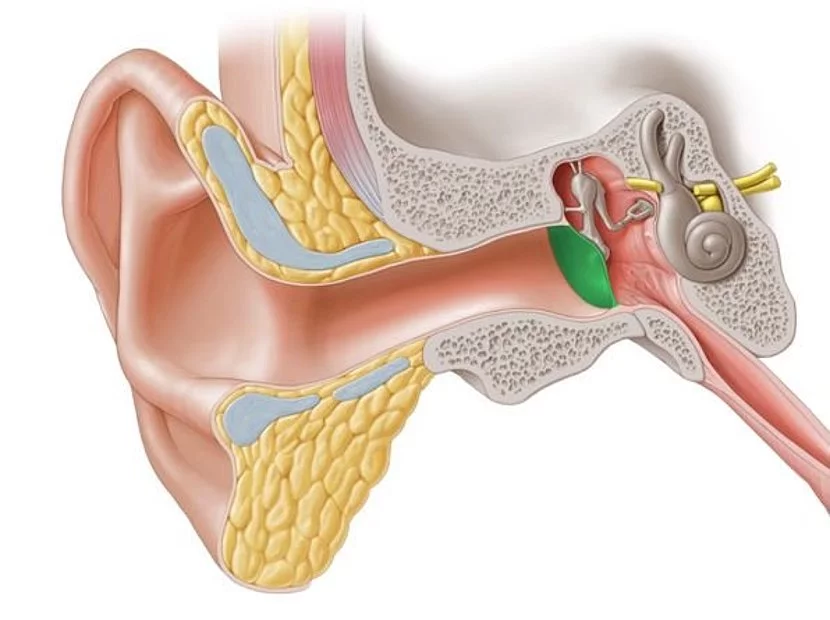
The petrous part of the temporal bones is among the most complex anatomical structures in the human body. Located directly behind the ears, the petrous part, houses the organs responsible for hearing and balance, as well as many nerves, such as the facial nerve, which supplies the facial muscles. All this is housed in a tiny space of just a few cubic centimeters, surrounded by the densest and hardest bone in the body.
Our recommendation
A detailed MRI or CT scan helps in the diagnosis of ear disorders and skull base pathologies.

Diseases of the temporal bones lead to pain in and deep in the ear, hearing loss, tinnitus, dizziness, balance disorders and nerve damage. Inflammation of the petrous part can spread to the brain and thus become life-threatening. The auditory and vestibular nerves and the facial nerve enter the temporal bones from the brain stem in the area of the inner connection between the temporal bones and the cranial cavity, the so-called cerebellopontine angle. Here, nerves and vessels that connect the vital brain stem are located in close proximity to each other. Even the smallest tumors, so-called acoustic neuromas, can cause pronounced symptoms. Early detection is crucial for successful treatment, particularly in this part of the body.
Our recommendation
Regular preventive care is important.
Adults up to the age of 40 should be screened every 4 years, and every 2 years after that.

High-resolution computed tomography (CT) can show the relevant structures of the petrous part in submillimeter resolution. This is essential for planning operations in the vicinity of the petrous bones.
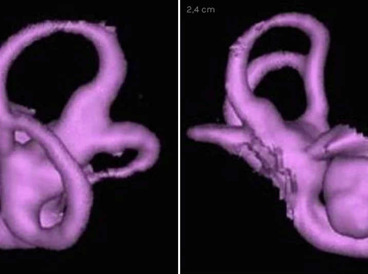
With magnetic resonance imaging (MRI), inflammations and tumors, i.e. soft tissue changes, can be detected precisely and at an early stage. In the case of hearing and balance disorders, i.e. hearing loss and vertigo, the endo- and perilymphatic structures, such as the cochlea and the semicircular canals of the inner ear, can be imaged in 3D with high detail resolution and examined for pathological changes using a special MRI examination.


