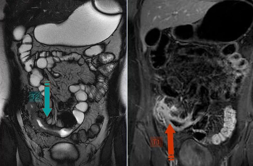
The small intestine is difficult to examine using conventional methods due to its location – in the middle of the gastrointestinal tract – and its length. MRI and CT enteroclysis examinations are special 3D examinations of the small intestine that enable the precise detection of inflammatory bowel diseases and tumors.
The MR enteroclysis examination is primarily used in younger people who are suspected of having Crohn’s disease or if complications (fistulas, stenoses, etc.) are to be assessed in cases where Crohn’s disease has already been diagnosed.
Risk factors
- Chronic inflammatory bowel disease (e.g. Crohn’s disease)
- Celiac disease
- Infections and parasites
- Lactose intolerance

MR enterography Crohn’s disease
Imaging of the entire small intestine. The normal intestinal wall appears without thickening and normal intestinal folds. The liver, gallbladder and stomach are visualized in the upper abdomen. Pathological intestinal loops with inflammatory wall thickening (blue and red arrow) are found in the right lower abdomen, diagnostic for Crohn’s disease.
CT enteroclysis is used for older people who need to be examined as quickly as possible due to their poor state of health and for whom radiation exposure is less important.


